Mr Lukasz Wojcik underwent ankle fusion (ankle arthrodesis) on 1 September 2026 under the care of Mr Peyman Bakhshayesh. Before surgery, he reported pain of 8 out of 10. At follow-up on 20 October 2026, exactly seven weeks after the operation, he reported zero pain.
- Before surgery
- 8/10
- At seven weeks
- 0/10
Preoperative pain
Reported on 20 October 2026
Pain scores and clinical details are reported by the treating surgeon from this individual patient’s experience.
The technique used
Mr Bakhshayesh used his preferred open ankle fusion technique through two incisions: one lateral and one anterior. The procedure involved:
- A fibular osteotomy through the lateral approach.
- Removal of the joint cartilage and preparation of the opposing bone surfaces, including feathering of the surfaces.
- Lateral fixation using a one-third tubular plate.
- A second plate placed anteriorly, creating orthogonal, dual-plate fixation.
In this approach, preparing the bone surfaces and providing fixation in two planes are intended to create a stable environment for fusion and support a planned programme of early weight bearing and rehabilitation.
Rehabilitation and early recovery
Mr Wojcik has already been referred for physiotherapy. At the seven-week review, his reported pain score was 0/10.
The timing and amount of weight bearing are decided for each patient, taking account of the fixation, bone quality, wound healing and follow-up assessment. Patients should follow their own surgeon’s instructions. Evidence about early weight bearing after ankle fusion is encouraging, but protocols vary and the quality of comparative evidence remains limited. [1]
Ankle fusion aims to relieve pain by joining the bones across the ankle joint. Movement at the fused joint is permanently lost, while other foot joints continue to contribute to walking. [2]
Before-and-after X-rays
The supplied images show the ankle before surgery and the plate-and-screw fixation after surgery. Select an image to open it at full size.
Before surgery
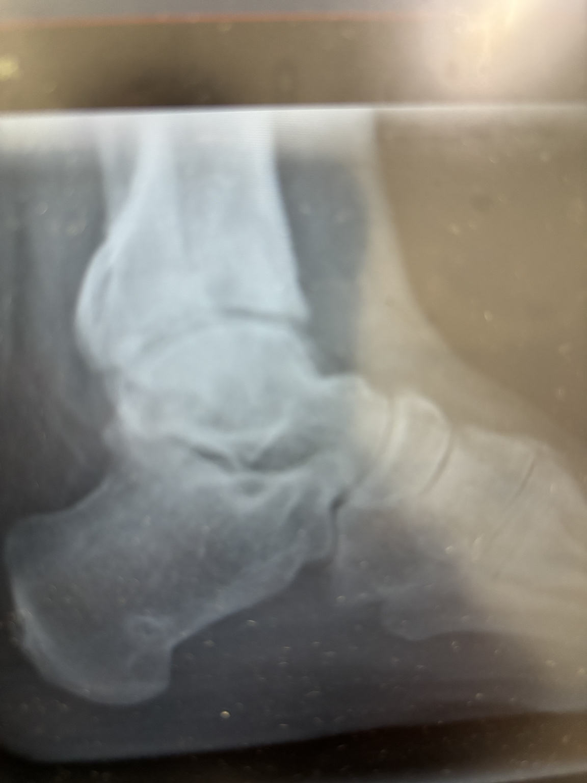
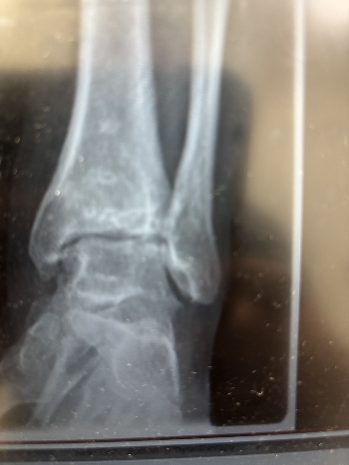
After surgery
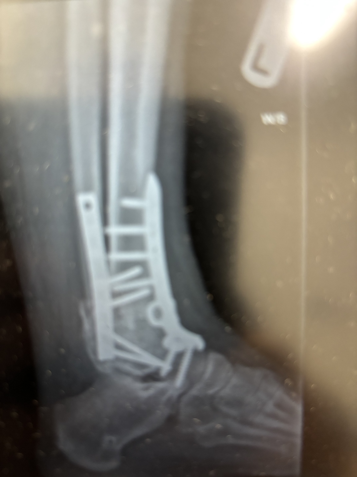
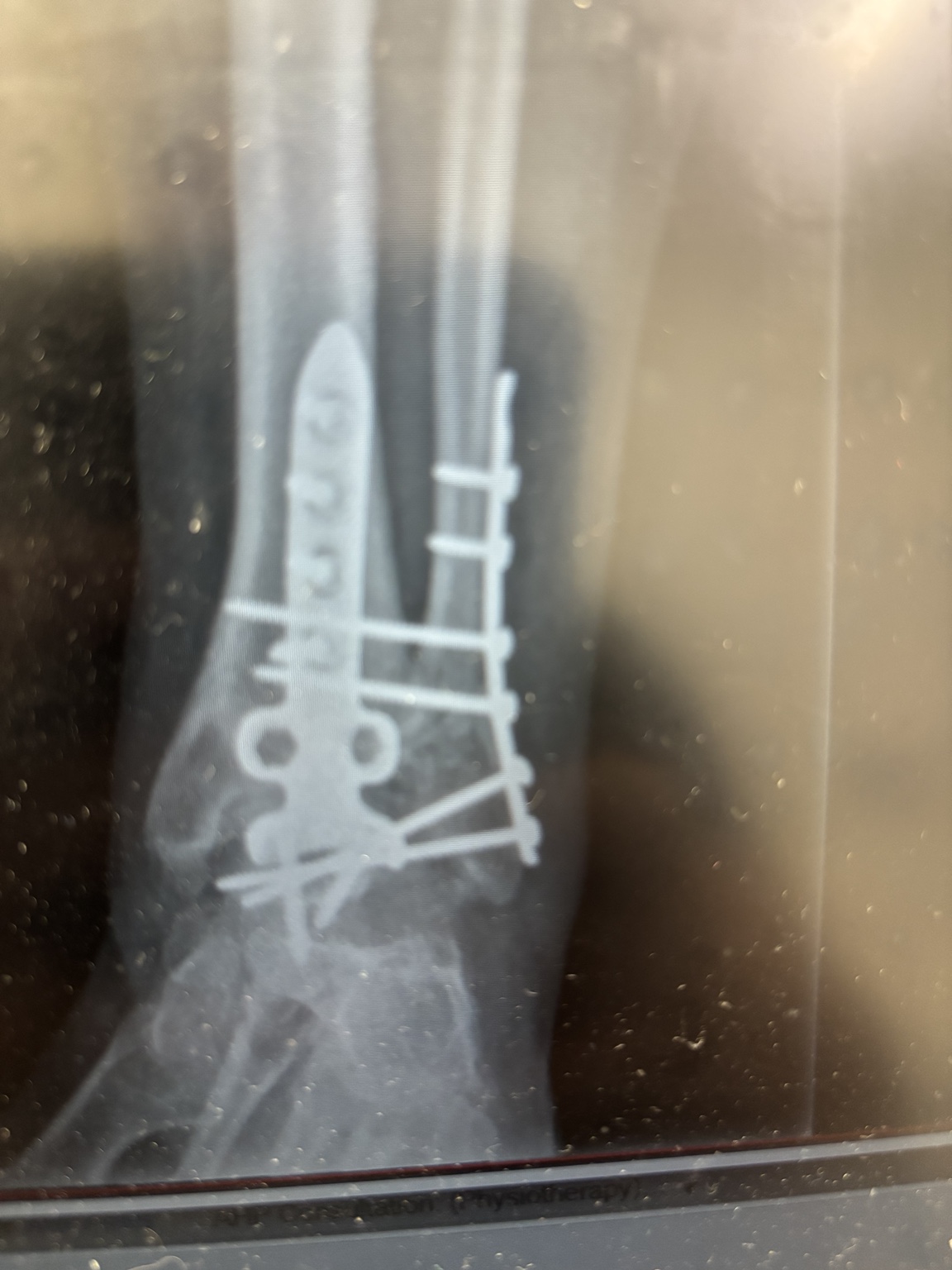
Original clinical images supplied by Mr Bakhshayesh. These photographs of X-rays are shown without changes to the anatomy or implants. The postoperative images show the fixation; assessment of fusion healing requires clinical review and follow-up imaging.
An early patient-reported outcome
This is a selected individual experience at seven weeks. It records Mr Wojcik’s reported pain relief at that point in recovery. Longer-term healing, function and rehabilitation continue to be assessed. Recovery and results vary between patients.
For treatment choices, risks and the usual recovery considerations, read the ankle fusion patient guide.
Further reading
- British Orthopaedic Foot & Ankle Society: weight-bearing after foot and ankle surgery
- Royal National Orthopaedic Hospital: ankle and hindfoot fusion patient guide
General clinical sources checked on 20 October 2026. The individual case details were supplied by the treating surgeon.