Patient information · Clubfoot · Flatfoot reconstruction
Flatfoot Reconstruction After Clubfoot
Patient information on combined calf lengthening, FDL tendon transfer, heel osteotomy and first tarsometatarsal fusion, including recovery, risks and postoperative X-ray examples.
Mr Peyman Bakhshayesh, MD, PhD, MBA
Consultant Trauma and Orthopaedic Surgeon
10 November 2026 · General patient information
An osteotomy and fusion need protection while bone and tendon heal. Follow your written weight-bearing instructions; the early walking advice for an isolated calf release does not apply automatically.
Rigid Flatfoot Following Childhood Clubfoot →
What does reconstruction aim to achieve?
Reconstruction aims to improve painful alignment and create a foot that meets the ground more evenly. It may help walking, shoe wear and stability, but cannot guarantee a normal-looking, fully flexible or pain-free foot. Treatment follows examination and standing imaging, with consideration of non-operative options.
The combination described here was selected by Mr Peyman Bakhshayesh for a rigid flatfoot following childhood clubfoot. Other patients may need different procedures, including additional fusion if a hindfoot joint is severely deformed, stiff or arthritic. These pages explain principles and the surgeon's account of this example; they do not prescribe the same operation for everyone. [1], [3]
1. Calf musculotendinous lengthening
The gastrocnemius and soleus are the calf muscles that feed into the Achilles tendon. Tightness can limit upward ankle movement and place additional strain on the reconstructed foot. Lengthening at the muscle–tendon region can reduce this pull. [8]
In this example, Mr Bakhshayesh describes a modified Strayer-type release involving the gastrocnemius musculotendinous region with additional soleus release. A standard Strayer procedure releases the gastrocnemius aponeurosis while leaving the soleus fascia intact; adding soleal fascial lengthening is a modification. This is distinct from lengthening the Achilles tendon farther down the leg. [4]
Nearby nerves, including the sural nerve, require protection. Possible consequences include numbness, calf weakness, altered calf contour and excessive lengthening. The amount and level of release are chosen according to the contracture. [8]
2. FDL-to-tibialis-posterior tendon transfer
The flexor digitorum longus (FDL) tendon normally helps bend the lesser toes. In the operation described, it was transferred to the tibialis posterior tendon to support the tendon mechanism along the inner side of the foot.
The transfer changes the direction of a tendon’s pull. It works alongside correction of the bones; a tendon transfer alone cannot reliably straighten a fixed bony deformity. Other muscles continue to help the toes bend, although some reduction in lesser-toe flexion strength is possible. Rehabilitation gradually trains the transferred tendon for its new role. [5], [9]
3. Heel-bone osteotomy and screw fixation
An osteotomy is a controlled cut through bone. The heel bone is repositioned to improve its relationship to the leg and the rest of the foot, then held while it heals. This helps redistribute loading and supports the soft-tissue reconstruction. [5]
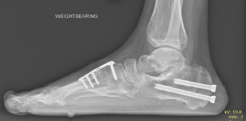
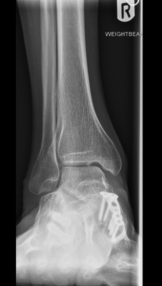
Mr Bakhshayesh reports fixation with 6.5 mm partially threaded Orthosolutions screws in this example. The screws hold the correction; they do not remove the need to protect the bone during healing. Prominent or uncomfortable screws occasionally require removal after the bone has united.
4. First tarsometatarsal fusion for forefoot supination
When the heel has been corrected, the inner forefoot may remain raised. The first tarsometatarsal (TMT) joint lies between the base of the first metatarsal and the medial cuneiform. Fusion can hold this part of the foot in a corrected position and provide stability where a limited fusion is appropriate. [6]
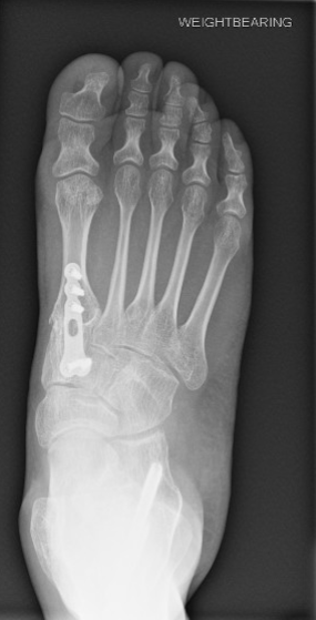
The joint surfaces are prepared and the bones held together while they unite. Mr Bakhshayesh reports using a four-hole Arthrex T-plate for the TMT fusion in this example. The aim was to address forefoot supination so the front of the foot could load more evenly. Fusion sacrifices movement at the treated joint; the choice depends on alignment, stability and joint condition.
Implant names describe the equipment used in this case. The most appropriate implant and procedure are selected individually.
Recovery and weight-bearing
The osteotomy, fusion and tendon transfer all need protection. Instructions for an isolated calf release do not apply automatically to this combined reconstruction. Your written discharge plan takes priority over the general timings below.
Initially, the foot is protected in a cast or splint. Elevation and keeping the dressing dry help wound recovery. Wounds are commonly reviewed at about two weeks. Mobility aids and practical arrangements at home are planned before discharge. [7]
- First six weeks: commonly no weight through the operated foot, using the prescribed walking aid. Some reconstructions require a longer period.
- Around six weeks onwards: examination and X-rays guide whether protected weight-bearing can begin. Do not increase loading simply because pain has settled.
- Around 8–12 weeks: cast or boot protection often continues through this period, and sometimes longer. Transition depends on bone healing and the surgeon's assessment. [6]
- Later recovery: physiotherapy, tendon retraining, strengthening and footwear progression are gradual. Swelling may persist for months; improvement can continue for a year or longer. [5]
- Work, sport and driving: timing depends on healing and demands. Driving requires safe vehicle control and an emergency stop, suitable footwear, no impairing medication and agreement with the treating team and insurer.
Risks and possible complications
Risks depend on the procedures, previous scars, bone quality, circulation, general health and healing. They are discussed individually; the list below is not a prediction of what will happen. Further treatment or surgery may be needed for a complication. [6], [7], [9]
- Infection, including deep infection involving bone or implants; bleeding or haematoma; delayed wound healing or wound breakdown.
- Abnormal, thickened, sensitive or painful scarring; numbness, tingling, nerve injury or persistent nerve pain; rarely, injury to blood vessels.
- Delayed union or non-union, meaning bone healing is slow or fails; malunion, meaning healing in an unsuitable position; loss of fixation or correction.
- Residual, recurrent, insufficient or excessive correction; stiffness, ongoing pain, prolonged swelling or continued difficulty with footwear and activity.
- Tendon-transfer stretching or failure, calf weakness, excessive lengthening, reduced push-off strength or reduced lesser-toe flexion strength.
- Prominent or painful metalwork, loosening or breakage of screws or plates, and possible later metalwork removal.
- Complex regional pain syndrome (CRPS): persistent pain with sensitivity, swelling, colour or temperature changes that may require specialist treatment.
- Deep vein thrombosis (DVT) or pulmonary embolism (PE): a clot in the leg or lung. Individual assessment determines whether blood-clot prevention medication is needed.
- Development or progression of arthritis in remaining joints, including adjacent-joint degeneration; post-traumatic arthritis where a joint has been injured.
- Anaesthetic complications and the possibility of future surgery, including revision fixation, repeat fusion or additional reconstruction.
Helping recovery and knowing when to seek help
Avoid smoking and nicotine, optimise diabetes control where relevant, eat adequately and follow the agreed wound, medication and loading instructions. Tell the team about previous blood clots, medication and any difficulty managing at home. Attend follow-up so changes to protection and activity reflect actual healing.
Seek urgent assessment for increasing calf pain or swelling, worsening wound redness, discharge, fever, severe escalating pain or cold/discoloured toes. Call 999 for sudden breathlessness, chest pain or collapse. Contact the treating team promptly if the cast feels too tight or you develop new persistent numbness. [7], [10]
Postoperative X-rays
These are postoperative weight-bearing X-rays supplied by Mr Bakhshayesh. They show the fixation used in this reconstruction. The calf release and tendon transfer are not directly visible on plain X-rays. Images alone do not establish bone union, pain relief or the long-term result.
Select an image to view it at full size.
Sources and further reading
- Faldini et al. Surgical management of clubfoot overcorrection: a case series (2023)
- Johnson et al. Late Effects of Clubfoot Deformity in Adolescent and Young Adult Patients Whose Initial Treatment Was an Extensive Soft-tissue Release: Topic Review and Clinical Case Series (2020)
- Eberhardt et al. Limitation of flatfoot surgery in overcorrected clubfeet after extensive surgery (2018)
- Firth et al. Lengthening of the gastrocnemius–soleus complex: JBJS supplementary surgical anatomy
- American Orthopaedic Foot & Ankle Society: Flatfoot surgical correction
- Royal Orthopaedic Hospital: Tarsometatarsal (midfoot) fusion
- Royal National Orthopaedic Hospital: Adult acquired flatfoot and tibialis posterior tendon dysfunction
- American Orthopaedic Foot & Ankle Society: Gastrocnemius release (Strayer procedure)
- Chelsea and Westminster Hospital NHS Foundation Trust: Treating the flat foot
- NHS: Deep vein thrombosis (DVT)
Published 10 November 2026. The case procedures and implant details are Mr Bakhshayesh’s supplied operative description. Sources on adult-acquired flatfoot explain general reconstruction principles; they do not define this post-clubfoot diagnosis or predict an individual outcome.
Arrange an assessment
Contact the practice · 07427 297799
Related guide: Rigid Flatfoot Following Childhood Clubfoot →
Calf and Achilles tightness · Complex regional pain syndrome